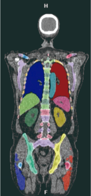
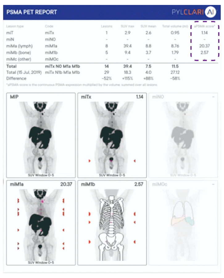
PYLCLARI AI®

The Al-Powered Assistant for Whole Body Automated PSMA Quantification and Standardized Reporting1
miT
miN
miMa
miMb
Source of PET/CT scan:
Patient data from the PYTHON study
PYLCLARI AI® is a web application that performs automatic segmentation and quantification of PSMA PET/CT images1

Quantitative

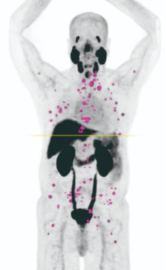
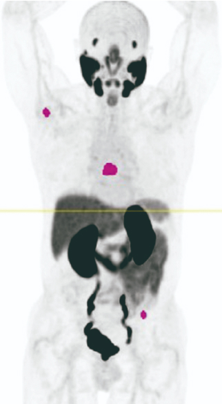
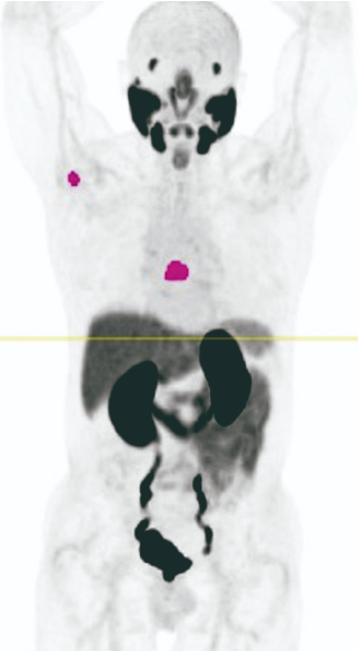
Assess all your patients' whole body PSMA PET/CT scans1
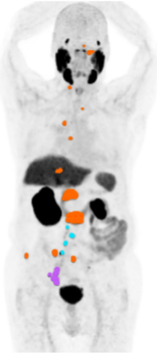
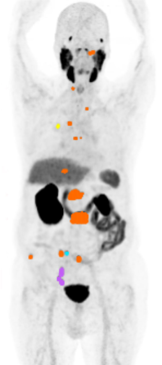
Reproducible

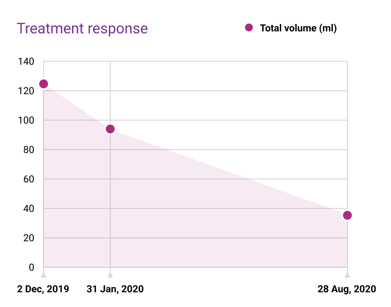
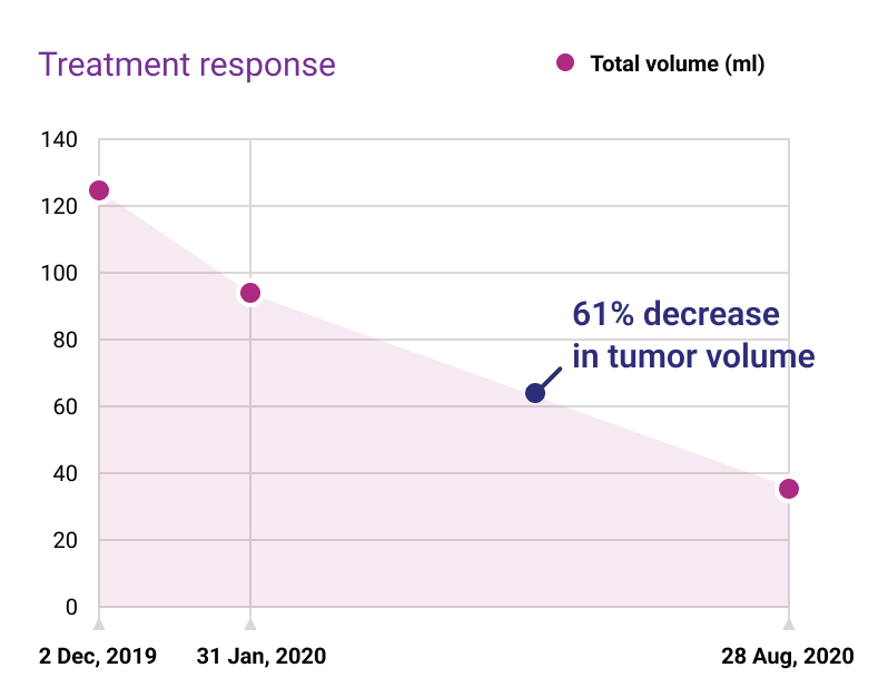
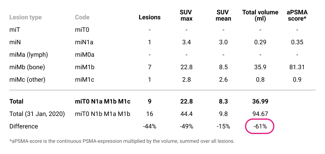
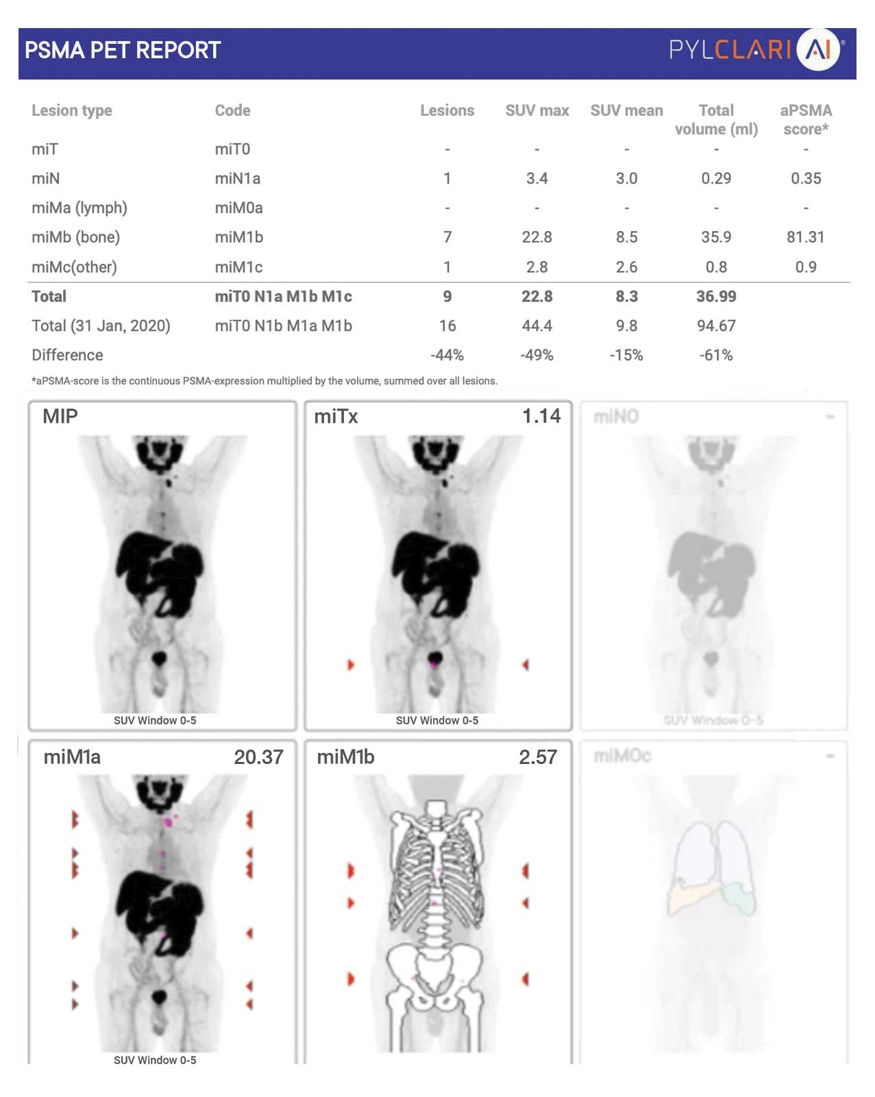
Create robust reports of patients' initial and follow-up scans1

Standardized

A single, comprehensive and standardized report1

AI-supported workflow

Automatically calculates organ segmentation and identifies hotspots1
CT component of
PSMA PET/CT
Al-based
CT segmentation of bone
and soft tissue
Al-based
Hotspot detection

and segmentation
Automated estimation of normal uptake (liver, blood)
Automated anatomical hotspot labeling (miTNM) and quantification
Physician review/validation
Quantitative outputs
Total PSMA SUVmean
Total PSMA tumor volume 

Total PSMA Score

Achieve higher reproducibility

98% when using PYLCLARI AI® against 90% for manual reads1
98%
ICC2
PYLCLARI AI®
90%
ICC2
Manual reads
PYLCLARI AI®
Manual reads
As assessed by Intraclass
Correlation Coefficient (ICC2) of three independent readers.

Compare patients' quantification results

For instance, total lesions volume, over time to assess disease progression or track treatment response1, 2
Baseline
miN
miMa
miMb
Month 2
miN
miMa
miMb
miMc
Month 10
miN
miMb
miMc

A single report

PYLCLARI AI® has a standardized report aligned to the PROMISE criteria1, 3, 4
Free text to be entered by reader for completing report
Estimation of physiological uptake
Automatic organ segmentation allows tumors to be classified by tissue type
Hotspots identified automatically
Patient and study information
The aPSMA score is a standardized measure of PSMA expression in prostate cancer lesions by integrating patient-specific PSMA-ligand uptake and the volume of each lesion 1,5
Lesion tissue type summary images
Compare patient results over time automatically
The PROMISE (Prostate Cancer Molecular Imaging Standardized Evaluation) criteria standardize the evaluation and reporting of PSMA PET/CT imaging in prostate cancer. Developed by experts in nuclear medicine, radiology, urology, and oncology, these criteria incorporate the miTNM classification, lesion characterization, and standardized metrics like the PSMA expression score to ensure consistent and accurate assessment of tumor burden across clinical settings.1,6

AI-supported workflow

Automatically calculates organ segmentation and identifies hotspots1
Sample report. Not an actual patient.
CT component of
PSMA PET/CT
Al-based
CT segmentation of bone and soft tissue
Al-based
Hotspot detection

and segmentation
Automated estimation of normal uptake (liver, blood)
Automated anatomical hotspot labeling (miTNM) and quantification
Physician review/validation
Quantitative outputsTotal PSMA SUVmean Total PSMA tumor volume 
Total PSMA Score

Achieve higher reproducibility

98% when using PYLCLARI AI® against 90% for manual reads1
98%
ICC2
PYLCLARI AI®
90%
ICC2
Manual reads
PYLCLARI AI®
Manual reads
As assessed by Intraclass
Correlation Coefficient (ICC2) of three independent readers.

Compare patients' quantification results

For instance, total lesions volume, over time to assess disease progression or track treatment response1, 2
Baseline
Month 6
Month 12

A single report

PYLCLARI AI® has a standardized report aligned to the PROMISE criteria1, 3, 4
Estimation of physiological uptake
The PROMISE (Prostate Cancer Molecular Imaging Standardized Evaluation) criteria standardize the evaluation and reporting of PSMA PET/CT imaging in prostate cancer. Developed by experts in nuclear medicine, radiology, urology, and oncology, these criteria incorporate the miTNM classification, lesion characterization, and standardized metrics like the PSMA expression score to ensure consistent and accurate assessment of tumor burden across clinical settings.1,6

Compare patients' quantification results

For instance, total lesions volume, over time to assess disease progression or track treatment response1, 2
Baseline
Month 6
Month 12

Achieve higher reproducibility

98% when using PYLCLARI AI® against 90% for manual reads1
98%
ICC2
PYLCLARI AI®
90%
ICC2
Manual reads
As assessed by Intraclass
Correlation Coefficient (ICC2) of three independent readers.

AI-supported workflow

Automatically calculates organ segmentation and identifies hotspots1
Sample report. Not an actual patient.
CT component of
PSMA PET/CT
Al-based
CT segmentation of bone and soft tissue
Al-based
Hotspot detection

and segmentation
Automated estimation of normal uptake (liver, blood)
Automated anatomical hotspot labeling (miTNM) and quantification
Physician review/validation
Quantitative outputsTotal PSMA SUVmean Total PSMA tumor volume 
Total PSMA Score
By clicking “Accept”, you agree to the storing of cookies on your device to enhance site navigation, analyze site usage, and assist in our marketing efforts. View our Terms of use for more information.